
Table of Contents
Ankle Sprain – Symptoms and causes
It is the most common acute injury in sports: you land on an opponent’s foot, catch an uneven edge on a trail, or misstep off a curb, forcing your ankle to roll violently inward. There is an audible pop, followed by an immediate flash of heat, sharp pain, and rapid swelling along the outer edge of your foot.
For most amateur athletes, the standard playbook involves limping to the couch, slapping a bag of frozen peas on the joint, popping a few ibuprofen, and waiting weeks for the swelling to go away.
Meanwhile, professional athletes sustaining the exact same injury are often back on the field or court in a fraction of the time. The secret to their accelerated recovery isn’t a miraculous genetic healing ability; it is a highly aggressive, scientifically validated medical protocol executed within the first 48 hours of the injury. While the average person treats a sprain with passive stagnation, sports medicine teams use targeted physiological management to protect tissue, control swelling, and jumpstart immediate cellular repair.
1. The Death of R.I.C.E.: Why Your Cold Pack Protocol is Outdated
For nearly four decades, the standard first-aid treatment for soft-tissue injuries was the R.I.C.E. protocol (Rest, Ice, Compression, Elevation). However, modern sports science has completely abandoned this framework because it actively delays tissue remodeling.
When a lateral ankle ligament—usually the Anterior Talofibular Ligament (ATFL) or Calcaneofibular Ligament (CFL)—is stretched or torn, your body triggers an inflammatory response. This inflammation isn’t a design flaw; it is a mandatory biological mechanism. Your body floods the area with white blood cells, growth factors, and signaling proteins needed to clean out damaged cells and lay down new collagen fibers.
  [Traditional R.I.C.E.] ──► Prolonged Freezing ──► Constricts Blood Vessels ──► Halts Inflammation ──► Stagnant Scar Tissue
  [Modern P.O.L.I.C.E.]  ──► Optimal Loading    ──► Cellular Mechanotransduction ──► Organized Collagen ──► Accelerated Return
When you wrap an ankle in ice for hours at a time, you drop the local tissue temperature so drastically that the surrounding blood vessels constrict. This vasoconstriction stops the inflammatory fluid from entering the zone, but it also completely halts the delivery of healing cells.
Worse yet, once the ice is removed, the local lymphatic system remains sluggish, trapping waste products and dead cells inside the joint. This creates a stagnant, fluid-logged environment that leads to thick, unorganized scar tissue and long-term joint stiffness.
2. Deciphering the P.O.L.I.C.E. Protocol
To accelerate recovery, professional sports medicine departments utilize the P.O.L.I.C.E. framework. This protocol focuses on protecting the joint while actively encouraging early, safe mechanical stress to speed up tissue regeneration.
1.Protection:Hours 0 to 4.
Immediately safeguard the joint using a rigid lace-up ankle brace or specialized structural taping. The goal is to completely eliminate lateral (side-to-side) twisting while still allowing the foot to gently bend up and down. Total immobilization in a stiff boot is avoided unless a fracture is suspected.
2.Optimal Loading:Hours 4 to 24.
Instead of complete rest, pro athletes introduce controlled mechanical stress to the tissue as early as possible. If a sports physician confirms the injury is a sprain and not a fracture, the athlete begins gentle, pain-free weight-bearing walks using crutches for support, or executes tiny, non-weight-bearing ankle circles.
3.Ice (For Pain Management Only):Hours 0 to 48 (Intermittent).
Ice is used strictly as a natural analgesic (pain reliever), never as a tool to compress or eliminate swelling. Apply cold packs for no more than 10 to 12 minutes at a time, allowing the tissue to return to normal temperature before reapplying. Stop using ice entirely after the first 24 to 48 hours.
4.Compression via a ‘U-Pad’:Hours 0 to 48 (Continuous).
Standard elastic wraps span smoothly across the ankle bones, leaving a hollow gap right underneath the lateral malleolus where fluid loves to pool. Pro athletes cut a “U-shaped” pad out of orthopedic foam, place it directly around the outer ankle bone, and wrap an elastic bandage over it to force fluid out of the joint recesses.
5.Elevation (True Venous Return):When Resting.
Propping your foot up on a coffee table isn’t enough. To actively drain metabolic waste from a swollen joint, the ankle must be positioned above the level of the heart. This creates a natural downhill slope, using gravity to channel fluid out of the lower leg and back into the central lymphatic system.
3. The Science of Optimal Loading: Mechanotransduction
The defining difference between amateur and professional injury management is the application of optimal loading. When soft tissue is subjected to mild, pain-free mechanical stress, the local cells undergo a biological process called mechanotransduction.
[Mild Mechanical Stress] ──► Activates Cellular Receptors ──► Stimulates Fibroblasts ──► Aligns Parallel Collagen Fibers
Inside your ligaments, specialized cells called fibroblasts are responsible for manufacturing new collagen strands to bridge the tear gap. If you remain completely stagnant on the couch, these fibroblasts lay down collagen in a chaotic, tangled, web-like pattern. This results in a weak, inelastic patch of scar tissue that is highly prone to re-tearing the moment you try to cut or sprint.
By introducing early, optimal loading—such as gentle heel-to-toe walking, seated calf stretches, or tracing the alphabet with your big toe—you generate tiny mechanical currents through the tissue. The fibroblasts detect these currents and lay down new collagen fibers in clean, organized, parallel lines that match the natural pull of the ligament. This ensures that when the ankle heals, it retains its original structural strength and elasticity.
4. Professional Modalities: Moving Beyond the Couch
While you are waiting out the clock, elite sports therapy rooms deploy advanced physiological tools within the first 48 hours to alter the tissue environment and accelerate the healing timeline.
                      ┌── Intermittent Pneumatic Compression (Mechanical pumping forces swelling out of joints)
Advanced Modalities ──┼── Neuromuscular Electrical Stimulation (NMES) (Pumps calf muscles to clear lymphatic fluids)
                      └── Kinesiology Floss Bands (Vascular restriction and rapid tissue re-perfusion)
Intermittent Pneumatic Compression (IPC)
Athletes regularly slide their legs into high-tech, full-length compression sleeves (such as Normatec boots). These devices utilize sequential, dynamic air pressure to mimic the body’s natural muscle pumps. The compression starts at the foot and cycles upward toward the hip, mechanically squeezing pooled inflammatory fluids out of the micro-spaces of the ankle joint and flushing them back into central circulation.
Neuromuscular Electrical Stimulation (NMES)
To drain an ankle without overloading the torn ligament, therapists place electrical stimulation pads directly onto the athlete’s calf muscles (gastrocnemius and soleus). The machine delivers low-voltage pulses that force the calf muscles to contract and relax rhythmically. This functions as an external “muscle pump,” driving lymphatic drainage and moving fluid upward out of the lower extremity without requiring the athlete to step or move the joint.
Tissue Flossing (Voodoo Flossing)
Therapists wrap a thick, heavy latex band tightly around the swollen ankle joint, starting below the heel and moving upward across the ankle bone. The athlete then performs basic, non-weight-bearing mobility drills for two minutes before the band is rapidly uncoiled. This temporarily restricts local blood flow, and the sudden release triggers a massive surge of fresh, oxygenated blood into the tissue, clearing out internal debris.
5. Critical Clearance: Sprain vs. High Ankle Sprain & Fracture
Before implementing any active mobilization or loading protocol, you must ensure that you are dealing with a standard lateral ankle sprain and not a far more severe structural injury.
[Inability to Take 4 Steps] OR [Bony Pinpoint Tenderness] ──► High Risk of Fracture ──► Order X-Ray (Ottawa Rules)
The Ottawa Ankle Rules
To rule out a bone fracture without rushing for an immediate X-ray, sports medicine clinicians apply the Ottawa Ankle Rules. If you meet any of the following criteria, stop loading the foot immediately and obtain radiological imaging:
- Inability to bear weight: You cannot take four consecutive steps immediately after the injury and during a physical assessment (even if limping).
- Malleolar bone tenderness: Pressing firmly along the posterior edge or tip of either the inner or outer ankle bone causes sharp, deep bone pain.
- Midfoot bone tenderness: Pressing directly on the base of the fifth metatarsal (outer midfoot) or the navicular bone (inner midfoot arch) causes sharp pain.
Detecting a High Ankle Sprain (Syndesmotic Tear)
A standard ankle sprain involves the lower lateral ligaments, but a high ankle sprain involves a tearing of the syndesmosis—the tough fibrous sheet holding your two lower leg bones (tibia and fibula) together. High ankle sprains require double the recovery time and must be completely immobilized early on.
To check for this, perform the Squeeze Test: take both hands and firmly squeeze your lower shin bones together midway up your calf. If this compression triggers sharp pain lower down, right above your ankle joint, the syndesmosis is likely damaged, and optimal loading protocols must be paused until you see an orthopedic specialist.
FAQ Section
Is it normal for my whole foot to turn purple after an ankle sprain?
Yes. Gravity naturally pulls blood and broken cellular debris downward into the lowest points of your extremity. Over the 3 to 5 days following a sprain, it is completely normal for deep purple, blue, and yellow bruising to spread all the way down into your midfoot, arch, and toes, even if the original injury was centered entirely on the ankle bone.
Can I take ibuprofen or naproxen right after a sprain?
Professional sports physicians generally advise against taking NSAIDs (Non-Steroidal Anti-Inflammatory Drugs) like ibuprofen, Advil, or Aleve during the first 48 hours. Because these medications chemically suppress the acute inflammatory phase, they disrupt the primary signaling cascade that triggers fibroblast recruitment and early collagen synthesis, potentially leading to a weaker long-term repair. Use acetaminophen (Tylenol) for early pain management instead.
How can I tell what grade my ankle sprain is?
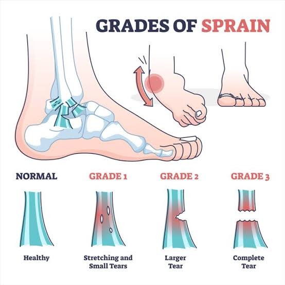
- Grade 1 (Mild): Micro-tearing of the ligament fibers. Presents with minimal swelling, mild tenderness, and an ability to bear weight almost immediately.
- Grade 2 (Moderate): A significant partial tear of the ligament. Characterized by moderate to severe localized swelling, visible bruising, limp-inducing weight-bearing pain, and mild joint laxity.
- Grade 3 (Severe): A complete rupture of the ligament. Features immediate, massive swelling, extensive deep bruising, an absolute inability to bear any weight, and a feeling that the joint is entirely unstable or sliding out of place.
Should I sleep with a compression bandage on my ankle?
You can wear a compression sleeve or elastic wrap to bed, but you must significantly reduce the tension before falling asleep. Your blood pressure drops naturally while sleeping, and a bandage wrapped too tightly can constrict local arterial flow, causing localized numbness, throbbing pain, or waking up with cold, pale toes.
When should I transition from a brace back to normal shoes?
You should begin weaning out of a supportive ankle brace once you can comfortably complete a normal heel-to-toe walking gait barefoot across a hard floor with zero pain or structural instability. Keep the brace handy for unpredictable surfaces (like grass, gravel, or crowded areas) until your single-leg balance matches the uninjured side.
Why does my ankle sprain keep re-occurring months later?
Repeated sprains occur due to a clinical deficit known as Functional Ankle Instability. When a ligament tears, the microscopic nerve receptors embedded inside that tissue (proprioceptors) are destroyed alongside the fibers. These nerves are responsible for telling your brain where your foot is in space.
If you do not perform specific balance and neurological rehabilitation drills after an injury, your brain fails to detect when your ankle is starting to tilt, preventing your peroneal muscles from firing fast enough to save the joint from rolling again.